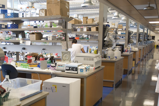
FACS Core Facility: Lymphocyte Analysis
Flow cytometry is a powerful technology for characterizing and analyzing cells as they move in a fluid stream through a beam of light. This high tech equipment enables researchers to count and characterize single immune cells from tens of thousands of cells circulating in the blood of patients, helping them understand how immune cells go awry in autoimmune diseases such as diabetes. Read more.
Histology Core Facility
The Histology Core Facility processes tissue samples for microscopic analysis. This core facility can process up to 300 issue samples at once, enabling low-cost and high-throughput microscopic examination of healthy and diseased tissue in the imaging core. Read more.
Imaging Core Facility
The Imaging Core Facility provides five workspaces equipped with powerful fluorescent microscopes for imaging both living cells and tissue obtained from healthy and diseased patients, as well as mouse models of disease. Read more.
Islet Isolation Core Facility
The Islet Isolation Core Facility provides islets (clusters of insulin-producing cells in the pancreas) from humans and mice for researchers in the Diabetes Research Program. These islets are an essential resource for our researchers studying aspects of islet biology, development, inflammation and transplantation. Isolation of islets of high yield and quality is technically challenging, and the expertise provided by the islet isolation core greatly enhances research productivity.
Viral Pathology Core Facility
The Viral Pathology Core Facility stores and grows viruses for diabetes research. In addition to being used to understand how viruses that infect insulin-producing beta cells can cause diabetes, these viruses can also be used for gene therapy approaches. This involves using viruses to transferring genes to beta cells to enhance their survival and function in diabetes, and after transplantation.