Projects
Current Projects
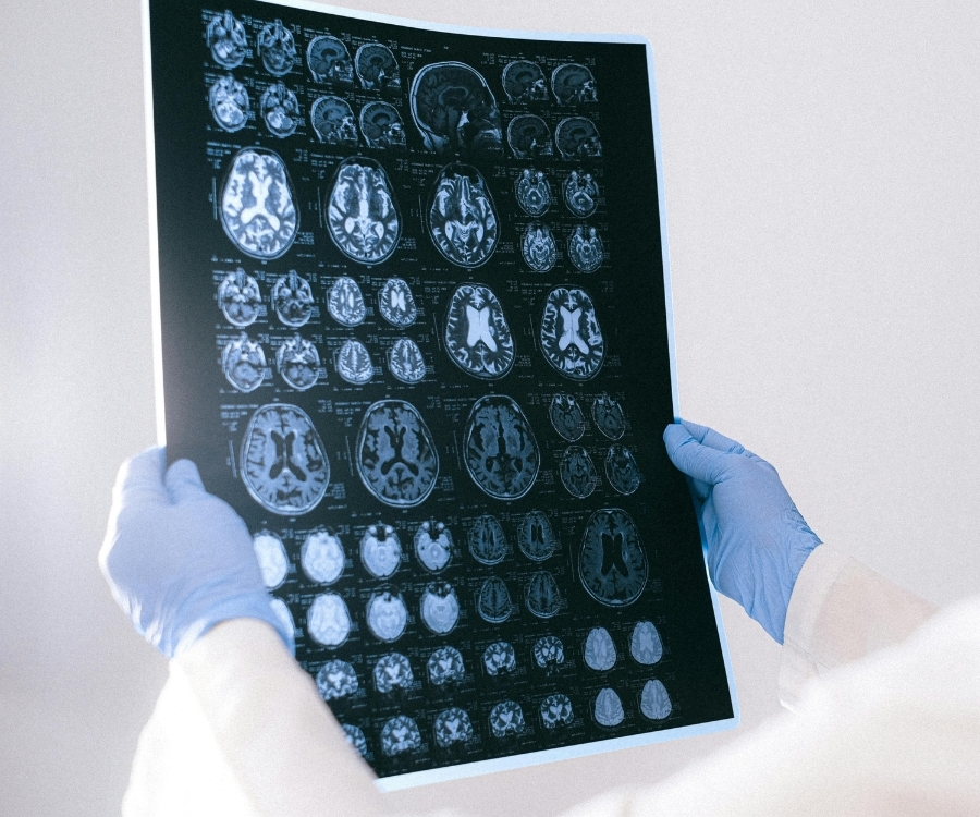
Investigating the Long-Term Memory of Functional Magnetic Resonance Imaging Time Series
Brain activity exhibits a scale-invariant nature: i.e. appears the same at varying levels of scale/magnification. This is shown in neuronal membrane potentials, neural field potentials, electroencephalography (EEG), magnetoencephalography (MEG), and fMRI, and is now seen as an integral feature of the brain. Scale-invariance is seen throughout nature, is a defining feature of fractal geometry, and is associated with long-range correlation in time. In the frequency domain, this is seen as a power-law decrease of the power spectrum, and is separate but coexists with brain oscillations.
Unlike brain oscillations, scale-invariant behaviour is not well understood. Research into this field of fMRI has revealed that analysis of the signal’s scale-invariant nature enables the discrimination of white matter and cerebrospinal fluid, active from inactive grey matter during a block paradigm, between rest and task, and neurological disorders. The scale-invariant phenomena of fractal signals can be completely characterized by two parameters: the variance and Hurst exponent (H). Typical Hurst values reported in the brain range between 0.5 to 1.0, with a common pattern of higher values dominating cortical regions, characterizing scale-invariant signals, and lower values seen subcortically. More persistent long-memory signals have been found to localize in the gray matter, with an average voxel-wise H of ~0.8, while white matter and cerebrospinal fluid tissue types are commonly defined by values closer to 0.5. As neuronal cell bodies reside in gray matter regions, scale-invariant temporal dynamics are suspected to originate neuronally and observed persistent scale invariance likely indicates synchronized neuronal oscillations rather than neurophysiological noise.
Scale-invariant analysis of fMRI BOLD signals remains relatively obscure, leaving much work to be done with regards to developing the best practices for analyses, in understanding and interpreting results, and the implications for neuroscience. This is where our research program aims to provide original and innovative contributions to the field.
Sucrose Therapy in Preterms: A Resting State fMRI Fractal Analysis Study
Preterm and critically ill newborns admitted to a NICU undergo repeated skin-breaking procedures that are necessary for their survival. In many parts of Canada, and North America, sucrose is becoming the accepted clinical standard nonpharmacologic intervention for managing acute procedural pain for these infants. Although shown to be safe in single doses, not enough studies have evaluated the effects of repeated doses of sucrose over relatively short periods of time. Furthermore, it is unclear how repeated doses of sucrose may alter functional brain development at such a critical period. Using data obtained in collaboration with Dr. Steven Miller at Sick Kids, we propose to use a novel resting state fMRI analysis method, known as Fractal Analysis, to study the fMRI brain dynamics of preterm infants who have received different doses of sucrose. This information could help us shed light on how sucrose may be altering brain signalling at a critical period of brain development.
Brain Health in Preterm Infants: Cerebral Metabolic Rate of Oxygen (CMRO2) Brain Mapping
Greater understanding of cerebral brain health in sick preterm infants is an important aim of neonatal medicine. Central to this premise is the development of quantitative, reliable, and non-invasive techniques for the assessment of cerebral circulation and metabolism. However, most current clinical techniques only measure the level of oxygen delivery, and not oxygen metabolism. Measures such as cerebral metabolic rate of oxygen (CMRO2), which provide information about the balance of cerebral oxygen delivery and demand, would have much greater clinical relevance. While there have been previous attempts to measure CMRO2 in newborns, we believe advanced magnetic resonance imaging (MRI) using a combination of quantitative susceptibility mapping (QSM) and arterial spin labelling (ASL) is potentially more accurate and precise, and ultimately more useful for studying and developing therapies for brain injured preterm and term neonates.
The overarching study aims are: use QSM and ASL to improve non-invasive measures of cerebral CMRO2 in very preterm infants. My lab will develop and improve our method of measuring CMRO2 in preterm children and establish their associations with clinical measures of health such as time on ventilation (ToV). This project has several phases: 1) improve protocol on scanner and collect pilot data from preterm neonates (24 to 32 weeks gestational age (GA); n = 20); 2) evaluate the relationship between CMRO2 and clinical risk factors; 3) Using data from Phases 1 and 2, conduct a larger study on very preterm (24 to 32 weeks GA; n > 50), moderately preterm (32 to 37 weeks GA; n > 50), and healthy term newborns (37 to 42 weeks GA; n > 50).
Developing these tools and innovations will place Canada and BC at the forefront of advanced quantitative MRI imaging of newborns. The resulting techniques will benefit future patients, healthcare providers, researchers, and the overall healthcare system.