Imaging Equipment
Molecular Devices ImageXpress Micro Confocal (IXMC)
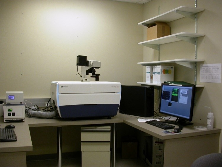 Molecular Devices ImageXpress Micro Confocal (IXMC) The Molecular Devices ImageXpress® Micro Confocal (IXMC) system is a powerful high-content imaging platform capable of widefield and confocal imaging for live or fixed samples, from whole organisms to intracellular events. With fast, high-resolution imaging and flexible analysis tools, it supports advanced 2D and 3D assay development and real-time data processing.
For more information:
Molecular Devices ImageXpress Micro Confocal (IXMC)
Olympus BX61
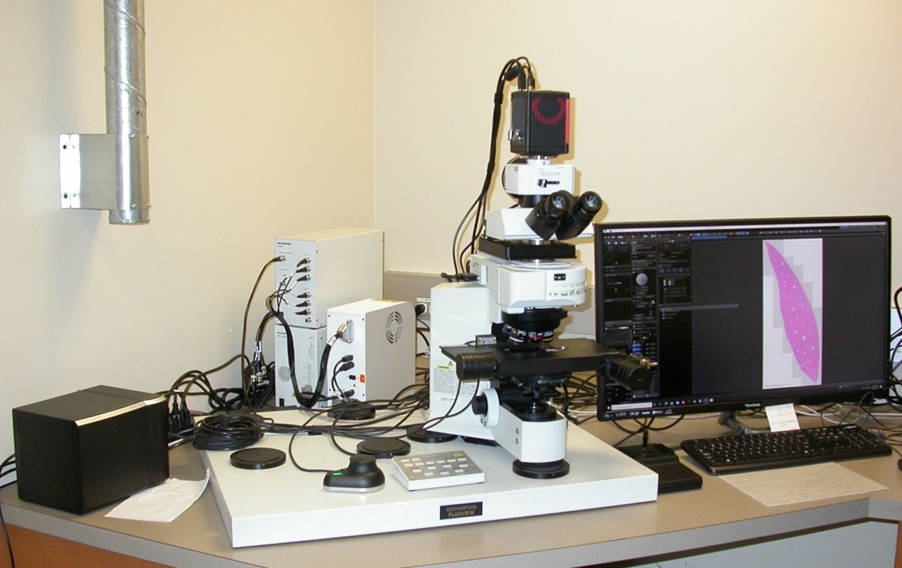 The Olympus BX61 is a versatile widefield microscope ideal for routine fluorescence and transmittance imaging, offering fast multi-channel tile scanning with real-time stitching and advanced 3D capabilities. Integrated with Olympus cellSens software, it delivers efficient image acquisition, processing, and analysis for a wide range of research needs.
For more information:
Comparison between Olympus BX61 and Keyence BZ-X810
Leica SP5II Laser Scanning Confocal & Wide Field Microscope
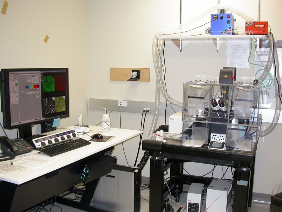 The Leica SP5II is a high-performance confocal and widefield imaging system offering flexible scan speeds, advanced spectral imaging, and exceptional sensitivity for live or fixed samples. With powerful features like hybrid detectors, 3D imaging, and an environmental chamber for time-lapse studies, it supports a wide range of sophisticated imaging applications.
For more information:
Leica SP5II Laser Scanning Confocal & Wide Field Microscope
Keyence BZ-X810 All-in-One Fluorescence & Bright Field Microscope
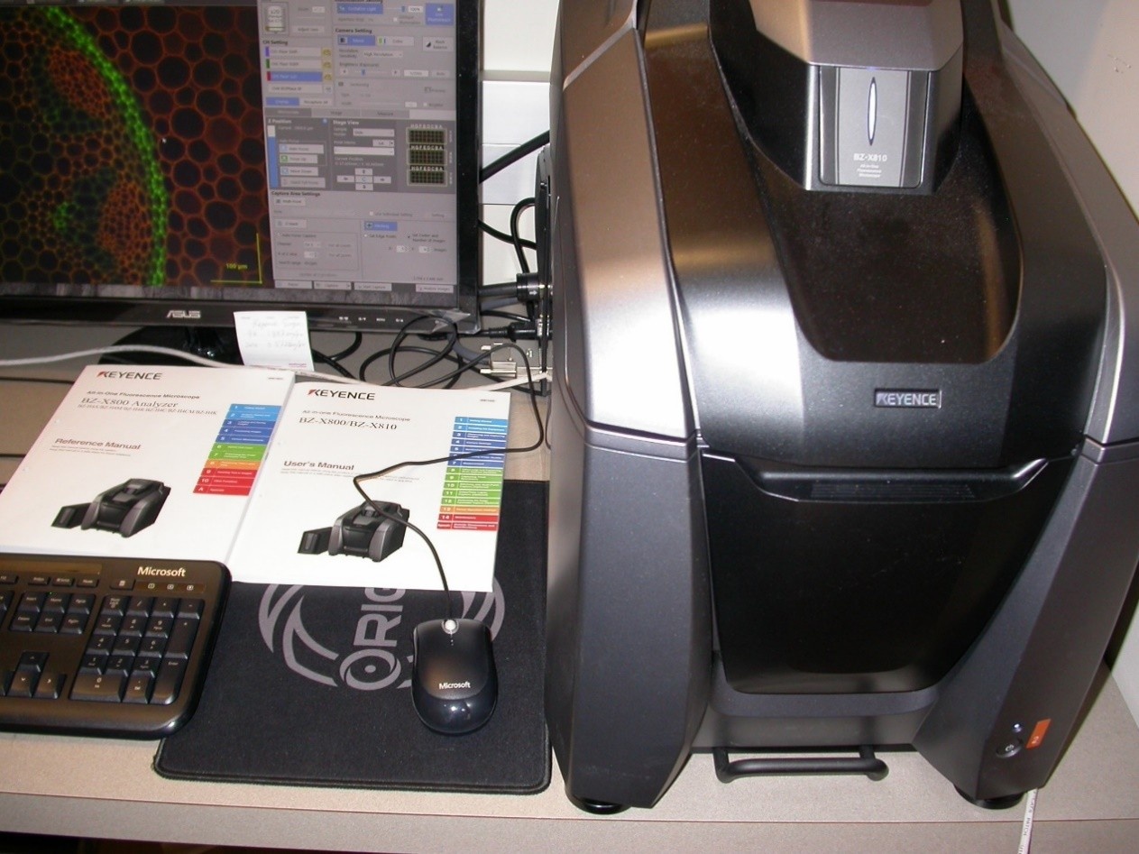 The Keyence BZ-X810 is an all-in-one fluorescence and brightfield microscope that combines high-speed, high-resolution imaging with intuitive automation for effortless slide scanning and image stitching. Its built-in darkroom, advanced autofocus, and easy operation make it ideal for capturing publication-quality images with minimal setup.
For more information:
Keyence BZ-X810 All-in-One Fluorescence & Bright Field Microscope
Comparison between Olympus BX61 and Keyence BZ-X810
Akoya PhenoCycler-Fusion 2.0 Multiplexing Spatial Biology Solution
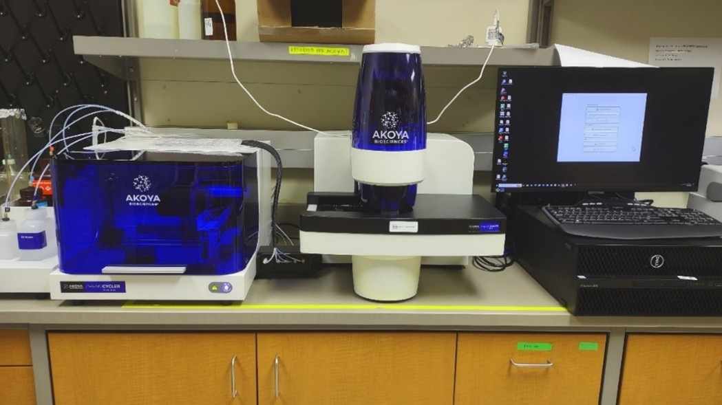 The Akoya PhenoCycler-Fusion 2.0 is a cutting-edge spatial biology platform that combines rapid, high-resolution imaging with automated multiplexing to analyze 100+ biomarkers across multiple tissue types. With parallel fluidics, iterative imaging, and oligonucleotide barcode technology, it delivers powerful insights into complex tissue architecture.
For more information:
Akoya PhenoCycler-Fusion 2.0 Multiplexing Spatial Biology Solution
Analysis Stations
High Content System Data Analysis Workstation 1 & 2
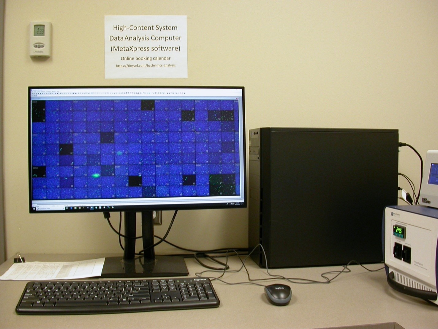 The High Content Data Analysis Workstations enable powerful, real-time image analysis even while imaging experiments are still in progress. Equipped with advanced application modules and a custom module editor, these identical offline stations support a wide range of high-content workflows—from cell scoring to neurite outgrowth analysis.
For more information:
High Content System Data Analysis Workstation 1 & 2
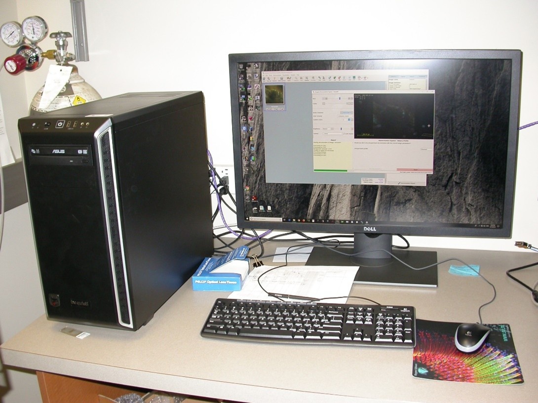
Image Deconvolution, 3D Rendering & Analysis Workstation
 This advanced workstation supports high-quality image restoration, 3D visualization, and batch deconvolution using Huygens Essential, Imaris, and other powerful tools. Ideal for enhancing confocal image clarity and generating realistic 3D renderings, it streamlines complex analysis with automated workflows and GPU acceleration.
For more information:
Image Deconvolution, 3D Rendering & Analysis Workstation
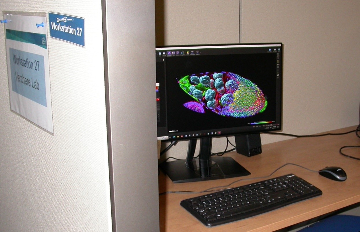
Imaris Image Analysis Workstation
Located at Workstation #27 in the shared office space, this powerful setup features the Imaris for Core Facilities package, enabling advanced 3D visualization, object detection, and quantitative analysis of widefield and confocal images. With GPU-accelerated deconvolution, smart workflows, and support for complex biological structures, it streamlines high-end microscopy data interpretation.
For more information:
Imaris Image Analysis Workstation
Guide to Microscope or Image Processing/Analysis Software Selection
How to select the proper microscope or Image Processing/Analysis software
Resources
Olympus BX61 Fluorescence and Bright Field Automated Upright Microscopes Specs
Leica SP5 Temperature Control Manual